10 Massages and Stretches for a Frozen Shoulder
June 15, 2023

Receiving an MRI report can feel like trying to read a foreign language. Words like "disc bulge," "herniation," "protrusion," and "degeneration" can sound alarming, often creating more anxiety than clarity. If you're holding a report right now, filled with technical terms and feeling a wave of concern, take a deep breath. You've come to the right place.
At Germanten Hospitals, we believe that an empowered patient is a healthier patient. Our philosophy, built on a foundation of German technological precision and decades of surgical expertise, starts with one simple thing: clear, honest communication. This guide is designed to do just that. We will walk you through your MRI report, demystify the jargon, and provide the crucial context you need to understand what these findings really mean for your health.
Think of us as your expert guide on this journey. We're here to turn confusion into confidence and fear into understanding, helping you take the first step toward a precise diagnosis and a life free from pain.
Before we can decode the report, let's quickly understand the part of your spine in question: the intervertebral disc. Your spine is made up of a series of bones called vertebrae, stacked one on top of the other. Between each of these bones lies a disc, which acts as a shock absorber and a flexible pivot point, allowing you to bend and twist.
The easiest way to understand a disc's structure is to think of a jelly donut. This simple analogy is used by spine specialists around the world because it's incredibly accurate. The disc has two main parts:
The health of your entire spine depends on this simple but brilliant design: a tough outer wall containing a soft, shock-absorbing core. Nearly all disc problems arise when this structure is compromised.
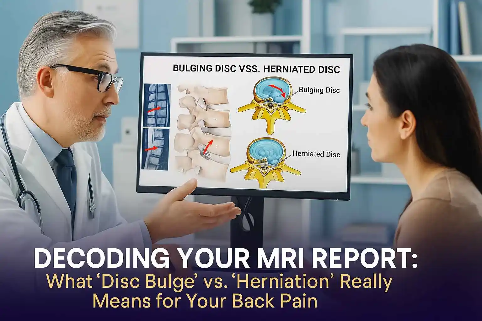
The terms "bulging disc" and "herniated disc" are often used interchangeably, but they describe two very different situations. The key distinction lies in the integrity of that tough outer wall, the annulus fibrosus.
A disc bulge is a condition where the disc sags and extends, or "balloons," outward from its normal space between the vertebrae. The most important thing to know about a bulge is that the outer wall (the annulus fibrosus) remains intact. There is no tear or rupture.
Radiologists, the specialists who read your MRI, define a bulge as a broad-based outpouching that involves more than a quarter (25%) of the disc's total circumference. This is why some experts use the analogy of a "hamburger that's too big for its bun". The entire disc is slightly flattened and sags outward evenly.
Disc bulges are extremely common and are often a normal part of the aging process, known as degenerative disc disease. As we age, our discs naturally lose water content and become less flexible. As the disc flattens, the annulus can lose tension and bulge outward, much like a car tire that has lost some air. In many cases, a bulging disc causes no symptoms at all.
A disc herniation is a more specific and often more significant event. Unlike a bulge, a herniation is defined by a tear or rupture in the annulus fibrosus. This tear creates a weak spot, allowing the soft, gel-like nucleus to push through the wall and leak out into the spinal canal.
This is why a herniation is often called a "ruptured disc." The term "slipped disc" is also commonly used, but it's a misnomer—the entire disc doesn't actually slip out of place. Radiologically, a herniation is a focal event, meaning it affects less than 25% of the disc's circumference.
Your MRI report might use even more specific terms to classify the type of herniation. Understanding these can give you a clearer picture of what the radiologist observed:
Here is a crucial point that is often overlooked: the severity of your pain is not always related to
the size of the herniation. A large disc bulge might cause no pain, while a tiny herniation can be
excruciating. Why?
The answer lies in biochemistry. While a bulge can cause pain by physically pressing on a nerve, a herniation adds a second, powerful pain-causing mechanism: chemical irritation. The material of the nucleus pulposus is recognized by your body's immune system as a highly inflammatory substance. When it leaks out and comes into direct contact with a nerve root, it triggers a severe inflammatory response. This chemical fire is a primary driver of the intense, burning, and radiating nerve pain known as sciatica. This is why treating the inflammation is just as important as addressing the mechanical pressure.
An MRI (Magnetic Resonance Imaging) scan is the gold standard for diagnosing issues with the soft tissues of your spine, like discs and nerves. Unlike an X-ray, which is excellent for looking at bones, an MRI provides a crystal-clear, detailed view of the discs, spinal cord, and nerve roots.
When one of our spine specialists at Germanten Hospitals reviews your MRI, they are performing a
systematic evaluation—a meticulous process of digital detective work. They aren't just looking for
one thing; they are assessing the entire ecosystem of your spine.
Here's what our experts are trained to look for:
Now we come to the single most important piece of information for any patient reading their MRI
report. This is a fact that can instantly reframe your perspective and alleviate a tremendous amount
of anxiety.
An MRI is a picture. It is an incredibly detailed and valuable picture, but it is not a diagnosis.
Decades of high-quality research have shown, without a doubt, that "abnormal" findings on spinal
MRIs are incredibly common in people who have absolutely no pain.
Many of the findings that sound so alarming on a report—disc degeneration, disc bulges, even disc
protrusions—are often just normal, age-related changes. They are the spinal equivalent of getting
wrinkles on your skin or grey hairs. They are a sign of time passing, not necessarily a sign of a
disease that needs fixing.
A landmark systematic review and meta-analysis published in the American Journal of Neuroradiology in 2015 compiled data from over 3,100 pain-free individuals to see what their spines looked like on an MRI. The results are eye-opening and provide powerful, reassuring context.
(Data synthesized from Brinjikji et al., AJNR, 2015)
Let this data sink in. If you are 40 years old and your report says you have a "disc bulge," you are
in the same boat as half of all other 40-year-olds walking around with no pain
at all. If you are 60 and your report mentions "disc degeneration," you share that trait with nearly
90% of your pain-free peers.
This doesn't mean your pain isn't real. It absolutely is. What it means is that the finding on the
MRI might just be an incidental finding—something that has been there for years without
causing a problem, and not the true source of your current pain.
This brings us to the heart of expert spine care. If the MRI picture alone can be misleading, what
truly determines your diagnosis? The answer is clinical
correlation.
At Germanten Hospitals, this is our guiding principle. An MRI report is just one tool in our diagnostic toolkit. The most important information comes from you. A diagnosis is only made when we can perfectly match the findings on your MRI scan to the specific symptoms you are experiencing.
This process of clinical correlation is a meticulous investigation that involves:
An expert spine specialist's job is to connect the dots. For example, if your MRI shows a herniation
at the L5-S1 level compressing the S1 nerve root on the right side, do your symptoms match? Do you
have pain radiating down the back of your right leg to your foot, and weakness when you try to stand
on your tiptoes? If the picture on the scan and the story your body is telling don't align, then the
finding on the MRI is likely not the culprit. This is how we identify the true "pain generator" and
avoid unnecessary treatments or surgeries.
Understanding your diagnosis is the first step. The next is understanding your path to recovery. And
here, there is more good news.
The natural history of most disc-related pain is overwhelmingly positive. The vast majority of patients—up to 90% in some studies—with a new disc herniation and sciatica will get significantly better on their own with conservative, non-surgical care.
Your body has a remarkable ability to heal. Over time, the inflammatory response subsides, and the
body can even reabsorb the herniated disc material, causing it to shrink and pull pressure off the
nerve. This process typically takes about 4 to 6 weeks for symptoms to improve significantly, though
full resolution can take a few months.
The goal of initial treatment is to manage the pain and inflammation while your body does its natural healing work. This typically includes:
Surgery is only considered when conservative treatments have failed to provide relief, or in the presence of specific "red flag" symptoms. These are signs of severe nerve compression that require urgent attention:
If surgery does become the right path for you, this is where the unique strengths of Germanten
Hospitals truly shine. Our entire approach is built on the principle of precision—using the most
advanced technology to solve the specific, confirmed problem with the least possible impact on your
body.
Long gone are the days of large incisions and lengthy, painful recoveries. Modern spine surgery is a story of micro-technology and precision engineering.
This combination of minimally invasive techniques and robotic precision ensures that we are targeting
only the source of the problem, preserving healthy tissue, and getting you back to your life faster
and more safely than ever before.
Your MRI report is a starting point, not a final destination. It is a valuable piece of data, but it
does not define you or your future. The most important takeaway is this: do not panic. The path to
recovery begins with a clear understanding of the true source of your pain, and that can only be
achieved through a comprehensive evaluation by a spine expert who knows how to connect the dots
between your imaging, your history, and your physical symptoms.
At Germanten Hospitals, our
world-class team, led by pioneers like Dr. Mir Jawad Zar
Khan, is dedicated to providing precisely that. We combine deep clinical
expertise with the most advanced diagnostic and surgical technologies to offer a level of care that
is both sophisticated and deeply personal.
Don't let an MRI report dictate your life. Let us help you decode it.
Schedule a comprehensive evaluation with the spine specialists at Germanten
Hospitals today. Let our experts provide the clarity you deserve and
create a personalized treatment plan to guide you back to a pain-free, active
life.
Q: Can a bulging disc heal on its own?
The symptoms from a bulging disc often resolve on their own over 6-8 weeks with conservative care.
While the bulge itself may not disappear, the pain and inflammation can subside, allowing you to
live symptom-free.
Q: Is a "bulging disc" the same as a "slipped disc"?
No. "Slipped disc" is
a common but inaccurate term for a disc herniation, where the inner material has actually ruptured
through the outer wall. A bulge is a less severe condition where the outer wall remains intact.
Q: How long does a herniated disc take to heal without surgery?
Most
patients feel significant improvement within 4 to 6 weeks. However, it can sometimes take several
months for the body to fully reabsorb the herniated material and for all symptoms to
resolve.
Q: What is the best sleeping position for a herniated disc?
Many people
find sleeping in the fetal position (on your side with your knees drawn up towards your chest) to be
the most comfortable, as it can open up the spaces between your vertebrae and relieve pressure on
the nerves. Placing a pillow between your knees can also help keep your spine aligned.
Q: How bad does my herniated disc have to be to need surgery?
The decision
for surgery is based less on the size of the herniation and more on your symptoms. Surgery is
typically considered only when you have persistent, debilitating pain that hasn't improved with
conservative care, or if you develop progressive neurological symptoms like significant weakness or
loss of bowel/bladder control.
Q: What is the success rate of microdiscectomy surgery?
For relieving
radicular pain (the pain that shoots down your leg or arm), microdiscectomy is a highly successful
procedure. Studies consistently show success rates in the range of 80-90% for significant pain
relief.