10 Massages and Stretches for a Frozen Shoulder
June 15, 2023
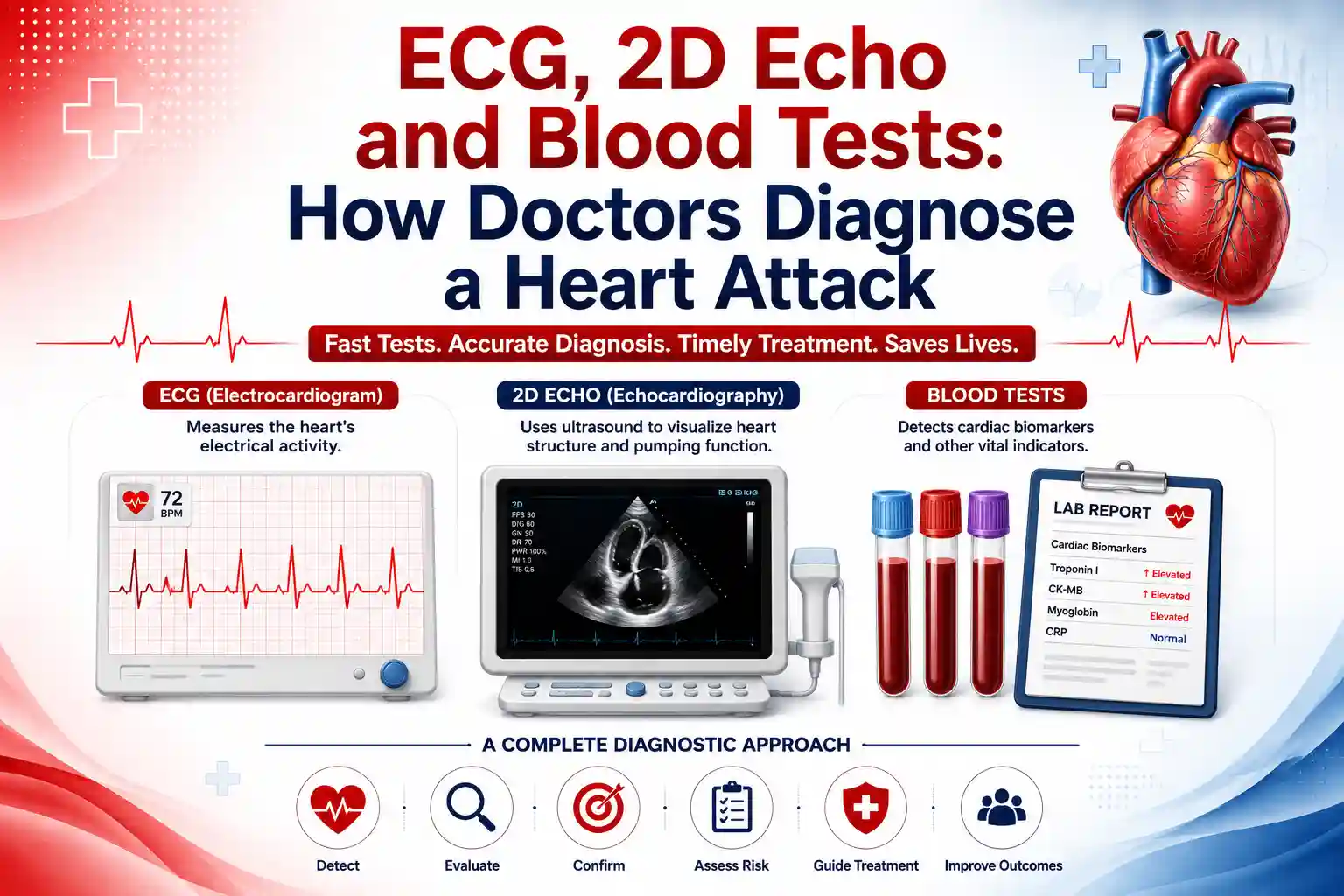
Chest pain brings a person to the emergency room. Within minutes, a nurse is attaching ECG leads to the chest. Soon after, blood is drawn for a test most people have never heard of. Sometimes a technician wheels in an ultrasound machine for an echo. Three different tests, all before anyone tells the patient what is actually wrong.
ECG vs 2D echo vs blood tests is a confusing comparison because doctors rarely use just one of these tools alone. Each test answers a different question about the heart, and diagnosing a heart attack accurately usually means combining all three. Understanding what each test does, and why none of them works alone, helps make sense of what happens in those first critical minutes.
A heart attack has two sides to it: an electrical side and a structural side, plus a chemical signature left behind in the blood. No single test captures all three. This is why doctors rely on ECG, 2D echo, and blood tests together rather than picking whichever one is fastest or most convenient.
Here is how the three tests compare at a glance:
| Test | What It Measures | Typical Time to Result |
|---|---|---|
| ECG | Electrical activity and heart rhythm | Minutes |
| 2D Echo | Heart structure, chamber size, wall motion, pumping strength | 20 to 40 minutes |
| Troponin blood test | Chemical evidence of heart muscle damage | About an hour, repeated a few hours later |
An electrocardiogram, or ECG, records the heart's electrical activity through electrodes placed on the chest, arms, and legs. A resting 12-lead ECG typically takes five to ten minutes from electrode placement to a completed reading, and it is usually the very first test ordered when someone arrives with chest pain, since it is fast, painless, and can immediately flag serious problems.
An ECG can show changes that point to a blocked artery or a prior heart attack, such as ST segment changes or Q waves. However, an ECG is only a snapshot of one moment in time. A normal ECG does not rule out a heart attack, especially early on or if the blockage has not yet caused enough electrical disturbance to appear on the trace. This is exactly why ECG results are read alongside symptoms and other tests rather than in isolation.
A 2D echocardiogram uses ultrasound waves to create moving images of the heart. Unlike an ECG, which only records electrical signals, a 2D echo shows how the heart muscle is actually moving, how well each chamber is pumping, and whether the valves are working properly.
For a heart attack, a 2D echo helps doctors see whether part of the heart wall is moving weakly or not at all, a sign that this area of the muscle was denied oxygen. It also helps estimate how much heart function has been affected, information an ECG cannot provide on its own.
When heart muscle cells are injured, they release a protein called troponin into the bloodstream. According to MedlinePlus, a troponin blood test measures how much of this protein is present, and it is one of the most important tools for confirming or ruling out a heart attack.
Troponin is usually tested more than once, often on arrival and again a few hours later, because levels rise as damage progresses. According to WebMD, modern high-sensitivity troponin tests can detect very small amounts of the protein and can help confirm a heart attack within about three hours of the first blood draw. A high troponin level strongly suggests heart damage, though doctors also consider other causes, since Cleveland Clinic notes that conditions such as kidney disease can raise troponin levels too.
Not every heart attack looks the same on these tests, which is part of why doctors run all three rather than stopping after the first one. In one type of heart attack, an artery is completely blocked, and this usually shows up clearly on the ECG within minutes, prompting immediate treatment to reopen the artery. In another type, the artery is only partially blocked, and the ECG may look normal or show only subtle changes, even though real damage is happening. In these partial-blockage cases, the troponin blood test often becomes the deciding factor, since it can detect heart muscle damage that the ECG alone missed.
This is also why a 2D echo carries extra weight when the ECG and early troponin results are unclear. Seeing a section of the heart wall move weakly gives doctors evidence of a real problem even before blood tests fully confirm it, which can be the difference between sending a patient home and admitting them for further care.
In practice, an ECG is done immediately on arrival, since it takes only minutes and can reveal an emergency needing instant treatment. Blood is drawn for troponin at the same time or shortly after, then repeated later to track any change. A 2D echo is often added when the diagnosis is uncertain, when doctors need to see how much of the heart muscle has been affected, or when planning further treatment.
None of these three tests is more important than the others. Together, they let a cardiologist confirm a heart attack accurately, rule it out when symptoms have another cause, and decide on the right treatment quickly.
Time matters in heart attack diagnosis. The sooner ECG, troponin, and echo results are available, the sooner treatment can begin, and the more heart muscle can potentially be saved. Access to cardiac tests in Hyderabad that combine same-day ECG, on-site lab testing for troponin, and echo imaging under one roof removes the delays that come from being sent between different facilities for each test.
ECG, 2D echo, and blood tests each answer a different question about the heart, and no single test tells the whole story. An ECG reveals the heart's electrical rhythm, a 2D echo shows its structure and movement, and troponin blood tests confirm whether the heart muscle has actually been damaged. Used together, they give doctors a fast, accurate answer during one of the most time-sensitive emergencies in medicine.
If you have unexplained chest pain, breathlessness, or risk factors for heart disease and want a thorough cardiac workup, book a cardiology appointment with Germanten Hospitals in Attapur, Hyderabad.
No. An ECG can show signs of a heart attack, but a normal ECG does not rule one out. Blood tests and sometimes a 2D echo are needed to confirm or exclude a heart attack completely.
Not reliably on its own. A 2D echo shows how well the heart muscle is moving, which can suggest damage, but it does not replace the electrical information from an ECG or the chemical confirmation from a troponin blood test.
Troponin levels rise gradually after heart muscle damage. Testing again a few hours after the first sample helps confirm whether levels are climbing, which supports a heart attack diagnosis, or staying flat, which points away from one.
Yes. Conditions such as kidney disease, heart failure, and other forms of heart strain can also raise troponin levels, which is why doctors interpret the result alongside symptoms and other tests rather than on its own.
A normal ECG does not rule out a heart attack, especially in the early hours. If chest pain persists, doctors typically still order troponin testing and monitor symptoms closely rather than dismissing the possibility based on the ECG alone.
A first ECG is usually available within minutes. Initial troponin results often come back within an hour, with a repeat test a few hours later. A 2D echo, when needed, can typically be completed within the same visit.