Month: July 2023

27 Jul 2023
What are the stages of ckd

What are the stages of CKD?

The progression of CKD varies for each individual and is influenced by factors like kidney function, underlying causes, and overall health. This article provides an informative overview of the stages of CKD, offering insights into the changes that occur as the disease advances. 

The 5 Stages of CKD: Tracking Kidney Function and Damage

Chronic Kidney Disease (CKD) is a long-term condition in which the kidneys gradually lose their ability to function correctly, leading to a decline in kidney function and potential complications. The stages of CKD are defined based on the estimated Glomerular Filtration Rate (eGFR), which is a measure of kidney function, and the presence of kidney damage. 

The 5 stages of CKD are as follows:GFR Dialysis

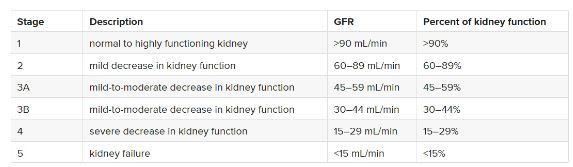
Stage 1: Kidney damage with normal or high eGFR 

  • eGFR > 90 mL/min/1.73 m²
  • Kidney damage is present, but kidney function is still considered normal.
  • Proteinuria (abnormal amounts of protein in the urine) may be detected.
  • Usually, there are no symptoms in this stage.

Stage 2: Kidney damage with mild decrease in eGFR 

  • eGFR 60-89 mL/min/1.73 m²
  • Kidney function is slightly impaired, but symptoms may still be absent. Fatigue
  • itching, loss of appetite, sleep problems and weakness are initial and nonspecific symptoms.
  • The kidneys are not working at full capacity, but the decreased function is mild.

Stage 3: Moderate decrease in eGFR

  • eGFR 30-59 mL/min/1.73 m²
  • Stage 3 is divided into two sub-stages:
  • Stage 3a: eGFR 45-59 mL/min/1.73 m²
  • Stage 3b: eGFR 30-44 mL/min/1.73 m²
  • Kidney function is significantly reduced, and the following symptoms may start appearing:
  • Back pain
  • Fatigue
  • Loss of appetite
  • Constant itching
  • Sleep problems
  • Hands and feet might swell
  • Too frequent or too rare urination
  • Weakness
  • Blood tests may show abnormalities related to kidney function.
  • Complications associated with reduced kidney function may arise:
  • Anaemia
  • Bone disease
  • High blood pressure

Stage 4: Severe decrease in eGFR 

  • eGFR 15-29 mL/min/1.73 m²
  • Kidney function is markedly reduced.
  • Symptoms and complications become more pronounced. 
  • While complications are the same as that in the case of stage 3 CKD, symptoms generally include: 
  • Back pain
  • Chest pain
  • Decreased mental sharpness
  • Fatigue
  • Loss of appetite
  • Muscle twitches/cramps
  • Nausea and vomiting
  • Persistent itching
  • Shortness of breath
  • Sleep problems
  • Hands and feet might swell
  • Too frequent or too rare urination
  • Weakness
  • Preparation for kidney replacement therapy (dialysis or transplantation) often begins at this stage.
  • At this stage the possibility of a heart disease or even a stroke increases.

Stage 5: End-stage kidney disease 

  • eGFR < 15 mL/min/1.73 m² or requiring dialysis
  • This is the most advanced stage of CKD.
  • Kidney function is severely impaired or completely lost.
  • Symptoms that are usually severe and can affect overall health include:
  • Back pain 
  • Chest pain
  • Breathing problems
  • Decreased mental sharpness
  • Fatigue
  • Little to zero appetite
  • Muscle twitches/cramps
  • Nausea or vomiting
  • Persistent itching
  • Trouble sleeping
  • Severe weakness
  • Hands and feet might swell
  • Too frequent or too rare urination
  • Dialysis or kidney transplantation is necessary to sustain life.

Watch this video to learn more about the five stages of chronic kidney disease in detail: 

Managing and Treating Chronic Kidney Disease: Key Approaches for Optimal Care 

Here are some key points about the treatment and management of CKD:

  • Blood pressure control: Maintain optimal blood pressure levels through lifestyle changes and medication to slow CKD progression.
  • Medications: Take prescribed medications to manage symptoms and complications like anaemia or mineral imbalances.
  • Kidney-friendly diet: Follow a diet that controls protein, sodium, potassium, and phosphorus intake to lessen kidney burden.
  • Fluid intake monitoring: Keep track of fluid intake to prevent fluid retention and complications.
  • Regular monitoring and tests: Attend regular check-ups, blood, and urine tests to monitor kidney function and adjust treatment plans.
  • Healthy lifestyle: Adopt a healthy lifestyle with regular exercise, weight management, no smoking, and limited alcohol intake slow CKD progression.
  • Manage underlying conditions: Treat and manage underlying conditions contributing to CKD like hypertension or autoimmune disorders, to minimise kidney damage.
  • Dialysis or transplantation: In advanced CKD stages, you must consider dialysis or kidney transplantation. 

Conclusion 

Remember, the progression of CKD can vary for each person, and proper management is crucial to slow down its progression and prevent complications. Regular monitoring and guidance from medical professionals are important for individuals with CKD.

FAQs

Q1. How long does it take to go from stage 3 to stage 4 kidney disease?

Ans. Most patients transition from stage 3 to stage 4 kidney disease in 10 years or above.

Q2. What foods are good for stage 5 CKD patients?

Ans. Fruits and vegetables contain less amount of potassium including grapes, green beans, cucumber, pasta, noodles and rice.

19 Jul 2023
signs and symptoms of kidney stones

Signs and Symptoms of Kidney Stones

Understanding the signs and symptoms can help identify and address kidney stones
promptly. This article provides an overview of kidney stones’ common signs and symptoms, helping you recognise potential indications and seek appropriate medical evaluation and treatment.

7 Common Signs and Symptoms of Kidney Stones: What to Look Out For 

Here is a list of 7 of the most common symptoms of kidney stones: 

1. Severe Pain

Kidney stones can cause excruciating pain, often described as colicky or cramp-like. Renal colic is one of the most hurtful kinds of pain that one can possibly imagine. The pain typically originates in the back, side, or lower abdomen.

Kidney stone pain usually begins suddenly and can change in location and intensity as the stone moves. The pain comes and goes in waves, which can be worsened by the ureter contracting to push the stone out. It is typically felt on the side and back below the ribs, and it may radiate to the belly and groin as the stone moves through the urinary tract. 

The size of the stone doesn’t always determine the severity of the pain; even small stones can be very painful when they move or cause a blockage.

2. Hematuria

The condition of the presence of blood in your urine is called hematuria. It is one of the most common and easily detectable symptoms of kidney stones. 

Blood may give the urine a pink, red, or brown colour. 

While sometimes this blood is easily visible when you are urinating, other times, the size of the blood cells may be extremely small to not be visible to the naked eye. In such cases, hematuria can be detected through a microscopic examination of the urine.

3. Nausea And Vomiting

Kidney stone pain can cause nausea and may lead to episodes of vomiting. This can occur due to severe pain or as a response to the body’s attempt to cope with the discomfort.

4. Urinary Urgency And Frequency

Kidney stones can cause an increased urge to urinate or a need to urinate more frequently than usual. 

Increased urgency and frequency of urination can signify that a kidney stone has moved into the lower part of the urinary tract. You may feel the need to use the bathroom more frequently throughout the day and night, sometimes even in a hurry. This symptom can be similar to the urgency experienced during a urinary tract infection (UTI).

However, the amount of urine passed may be minimal due to the blockage or obstruction caused by the kidney stone.

5. Painful Urination (Dysuria)

Some individuals with kidney stones may experience sharp pain or a burning sensation during urination. 

When a kidney stone reaches the point where the ureter and bladder meet, you may start experiencing pain while urinating, which is called dysuria. Discomfort can occur when the stone irritates the lining of the urinary tract as it passes through. 

If you’re unaware of the presence of a kidney stone, you might confuse the symptoms with a urinary tract infection (UTI). In some cases, there can be an infection along with the presence of the stone.

6. Cloudy Or Foul-Smelling Urine

Kidney stones can sometimes result in changes in urine appearance.  Cloudy or foul-smelling urine can indicate a urinary tract infection (UTI) or kidney infection. About 16% of people with acute kidney stones also have a UTI. As a result, the urine may appear cloudy or have an unpleasant odour.

Cloudiness suggests the presence of pus in the urine, while the smell may come from bacteria causing the infection. Strong odours can also result from concentrated urine. If a UTI occurs alongside a kidney stone, it is considered a surgical emergency, even if there is no fever.

7. Fever And Chills 

In some cases, kidney stones can lead to UTIs or kidney infections. Symptoms such as fever, chills, and general malaise may be present in such situations.

Conclusion

The presence and severity of these symptoms can vary from person to person. Additionally, some individuals with kidney stones may not experience any symptoms until the stone causes an obstruction or moves within the urinary tract. If you suspect kidney stones based on these signs, it’s important to seek medical evaluation for a proper diagnosis and treatment.


FAQs

Q1. What is the first symptom of kidney stones?

Ans. The first symptom of kidney stones can vary among individuals, but one common early symptom is intense pain. This pain often starts suddenly and can be felt in the back, side, or lower abdomen.

Q2. What does kidney stone pain feel like in a woman?

Ans. Kidney stone pain in women is often described as intense, sharp, and colicky.  The sensation is often compared to being stabbed with a knife or experiencing severe cramps.


12 Jul 2023
What is pediatric kidney diseas

What Is Pediatric Kidney Disease?

What Is Pediatric Kidney Disease?

‘Pediatric kidney disease’ is a broad-spectrum term that covers a range of conditions affecting the kidneys in children. This article includes a detailed guide on its causes, types, diagnosis, symptoms, and the role of healthcare professionals in treating them. 

Types of Pediatric Kidney Diseases

Various types of pediatric kidney diseases can affect children. Here are some common types: 

  1. Congenital Anomalies
  • Congenital Hydronephrosis
  • Polycystic Kidney Disease (PKD)
  1. Glomerular Diseases
  2. Inherited Disorders
  • Alport Syndrome
  • Nephronophthisis
  1. Urinary Tract Infections (UTIs)
  2. Renal Tubular Disorders
  3. Acquired Kidney Diseases
  • Acute Glomerulonephritis
  • Hemolytic Uremic Syndrome

Pediatric CKD (chronic kidney disease)

Kidney diseases are relatively uncommon in children. Many children may not exhibit noticeable symptoms until the disease has progressed. As a result, it becomes difficult to recognize the true number of affected children. 

Pediatric CKD is one of the most commonly occurring types of kidney disorders in [1] boys over the age of 6 years when compared to girls.

The disease results in long-term kidney damage or kidney dysfunction. While complete recovery is not possible, your healthcare professional can help you and your child to manage it effectively for a healthy lifestyle. 

Pediatric CKD progresses in 5 stages and is dependent on GFR (i.e. glomerular filtration rate).

Pediatric Renal Tumors

Pediatric renal tumours refer to the development of abnormal growths or tumours in the kidneys of children. They are relatively rare but represent a significant proportion of childhood malignancies. 

The most common types of renal tumours in children include—

  • Wilms tumour
  • Clear cell sarcoma of the kidney
  • Renal cell carcinoma 

Wilms tumour (Nephroblastoma):

Wilms tumour is the most common renal tumour in children. It usually affects children between the ages of 2 and 5. Imaging studies like ultrasound, CT scan, and MRI are used to evaluate the tumour and determine its extent.

Treatment typically involves a combination of surgery, chemotherapy, and sometimes radiation therapy. 

Clear Cell Sarcoma Of The Kidney (CCSK):

Clear cell sarcoma of the kidney is a rare malignant tumour that primarily affects young children. Treatment typically involves surgical resection of the tumour, sometimes followed by chemotherapy or radiation therapy depending on the specific case.

Renal Cell Carcinoma (RCC):

RCC is extremely rare in children.

Children with RCC often present with symptoms such as:

  • abdominal pain
  • hematuria (blood in urine)
  • or a palpable mass.

Treatment for pediatric RCC usually involves surgical removal of the tumour, followed by additional therapies if needed.

Imaging studies, such as ultrasound, CT scan, and MRI, help evaluate the tumour and determine its characteristics in all the above 3 cases. 

Pediatric Acute Kidney Injury

Pediatric acute kidney injury (AKI) refers to a sudden and sharp decline in kidney function in children. It is a serious condition that requires prompt recognition and the right actions to be taken at the right time to improve it. 

AKI can result from various causes, including dehydration, sepsis, medication toxicity, urinary tract obstruction, and certain medical procedures. 

Treatment may involve fluid resuscitation , optimizing hemodynamics , addressing the underlying cause, and providing renal support as necessary. 

Acute Renal Failure In Pediatrics

Renal failure refers to the inability of the kidneys to adequately filter waste products and maintain fluid and electrolyte balance. It can be acute or chronic, and it can result from various underlying causes or certain medications. 

Common symptoms may include—

  • Decreased Urine Output
  • Fluid Retention
  • High BP
  • Fatigue
  • Abnormal Electrolyte Levels 

Common Symptoms Of Kidney Disease In Children 

Most pediatric kidney diseases show little to no symptoms in the early stages and that is why their detection is not possible. However, when the disease gets worse following general symptoms are seen:

  • oedema
  • a significant increase or decrease in the production of urine. Some children might have the need to urinate more frequently sometimes resulting in wetting the bed.
  • proteinuria
  • hematuria
  • decreased appetite
  • feeling tired
  • fever
  • high blood pressure
  • itchy skin
  • nausea 
  • vomiting
  • shortness of breath
  • trouble concentrating
  • weakness
  • weight loss
  • stunted growth 

Diagnosis Of Pediatric Kidney Disease

The diagnosis of Children’s kidney diseases like pediatric renal tumours, pediatric acute kidney injury, acute renal failure in pediatrics, etc. is done using two types of methods: 

  • Non-invasive Methods
  • Invasive Methods

The non-invasive methods include:

  1. Physical Exam 
  2. Medical History
  3. Urinalysis
  4. Microalbuminuria
  5. Creatinine Clearance 
  6. Imaging studies
  • Standard X-rays 
  • Angiography 
  1. Intravenous Urography
  2. Ultrasounds
  3. CT Scans 
  4. Magnetic resonance imagery (MRI)

The invasive methods: 

  1. Blood Tests
  2. Serum creatinine 
  3. Blood Urea Nitrogen 
  4. GFR
  5. Voiding cystourethrogram (VCUG)
  6. Kidney Biopsy 

Treatment Approaches for Pediatric Kidney Disease

In order to treat and manage pediatric kidney diseases with maximum efficiency, it is very important to consider both: a pediatrician & a nephrologist. 

Your healthcare professional might start by prescribing antibiotics (in case of an infection) to treat the underlying cause of the specific type of disease. 

Treatment options depend on the type and severity of the kidney disease. They may include:

 Medications

Doctors may prescribe medications to control blood pressure, reduce inflammation, manage proteinuria (excessive protein in urine), or treat underlying causes such as infections or autoimmune conditions.

 Dietary Modifications

A balanced diet with appropriate restriction of certain nutrients, such as sodium and potassium, may be recommended to manage pediatric kidney disease. 

Dialysis

In cases of advanced kidney failure, dialysis may be necessary to filter waste products and excess fluid from the blood. Pediatric patients may undergo either hemodialysis or peritoneal dialysis, depending on individual circumstances.

Kidney Transplant

When kidney function is severely compromised, a kidney transplant may be considered. Transplantation offers the potential for long-term improvement in kidney function, providing a better quality of life for children with end-stage renal disease.

In addition to these treatments, healthcare professionals will also suggest regular monitoring, follow-up visits, and strict commitment to prescribed medications and lifestyle changes.

They work closely with pediatric nephrologists to develop a detailed care plan that addresses the unique needs of each child. 

Conclusion

All in all, when it comes to keeping your child healthy, the more you delay, the more complicated the disease gets. Therefore, it is essential to call the doctor at the right time regardless of whether your child is an infant or older.


04 Jul 2023
Can pulmonary oedema be treated?

Can Pulmonary edema Be Treated?

What is pulmonary edema?

Pulmonary oedema is a medical condition that is treatable. It occurs when there is an excess buildup of fluid in the air sacs of the lungs. 

This fluid buildup can make breathing difficult and cause symptoms such as coughing up foam or loose mucus, chest tightness, shortness of breath, and wheezing. 

Pulmonary oedema can be caused by various factors such as heart problems, pneumonia, toxin exposure, medications, or chest trauma.

It is of two types: cardiogenic and noncardiogenic. A heart problem causes cardiogenic pulmonary oedema, while other factors cause non-cardiogenic pulmonary edema. Pulmonary edema can be acute or chronic and life-threatening, making immediate treatment necessary.

Diagnosis of pulmonary edema

Diagnosing pulmonary oedema typically involves a combination of physical examination, diagnostic tests, and imaging studies. 

The following are some of the common diagnostic methods used to diagnose pulmonary edema:

  • Physical examination: A healthcare provider will perform a physical examination to check for signs of fluid buildup in the lungs. This may include listening to the lungs with a stethoscope to detect abnormal sounds such as crackles or wheezes.
  • Chest X-ray: A chest X-ray can help visualise the presence of fluid in the lungs, which can confirm a diagnosis of pulmonary oedema. It is one of the first tests ordered.
  •  Pulse oximetry: A small device clips onto a finger or earlobe and measures the amount of oxygen in the blood. This test can help determine if the lungs are effectively oxygenating the blood.
  • B-type natriuretic peptide (BNP) blood test: A BNP blood test measures the hormone level produced in response to excess fluid buildup by the heart. It can help distinguish between cardiac and non-cardiac causes of pulmonary oedema.
  • Chest computerised tomography (CT) scan: A chest CT scan is a series of X-ray images that creates a detailed picture of the lungs and can help identify the underlying cause of pulmonary edema.
  • Arterial blood gas test: This test measures oxygen and carbon dioxide level in the blood and can help assess the severity of pulmonary oedema.
  • Electrocardiogram: An electrocardiogram (ECG) is a test that records the heart’s electrical activity. This test can help diagnose any underlying heart problems causing pulmonary edema.
  • Echocardiogram: It is an imaging test that uses ultrasound to create detailed images of the heart. It helps to identify any abnormalities in heart function contributing to pulmonary edema.
  • Cardiac catheterisation: In this test, a thin, flexible tube is inserted into a blood vessel and threaded up to the heart. This test can help measure the pressure inside the heart and lungs, which can help diagnose pulmonary oedema.
  • Complete Blood Count: A complete blood count (CBC) test is done to 

identify any infections or other conditions contributing to pulmonary edema.

  • Lungs Ultrasound: A lung ultrasound is non-invasive and creates lung images using high-frequency sound waves. It is used to identify the presence of fluid in the lungs.

A timely and appropriate diagnosis is crucial for effectively treating and managing this potentially life-threatening condition.

Can pulmonary edema be treated? 

The treatment for pulmonary oedema is available and it depends on the underlying cause and the severity of the condition. The following are some common treatment options:

  • Medications: Diuretics are commonly used to help remove excess fluid from the lungs and improve breathing. Inotropes are used to help improve heart function. Morphine may be given to reduce anxiety and improve breathing, and blood pressure drugs can be used to reduce the workload on the heart.
  • Breathing Tube in the Trachea: In severe cases of pulmonary edema, a breathing tube may be inserted into the trachea that helps patients breathe more effectively.
  • Oxygen Therapy: Oxygen therapy may be provided using a mask, nasal cannula, or positive pressure mask to help increase the amount of oxygen in the blood.
  • Renal Replacement Therapy: In some cases, pulmonary edema may be caused by kidney failure. In such cases, renal replacement therapy, such as dialysis, may be necessary to remove excess fluid from the body.
  • Continuous Positive Airway Pressure (CPAP): CPAP therapy involves using a machine that delivers a continuous flow of air pressure to the lungs through a mask. It helps reduce the amount of fluid in the lungs and improve breathing.

In addition to these treatments, addressing the underlying cause of pulmonary edema is important. This may involve treating an underlying heart condition, such as heart failure or arrhythmia, or addressing an infection or toxin exposure.

Prevention of pulmonary edema

Here are some preventative measures you can take to reduce your risk of developing pulmonary edema:

  • Manage Blood Pressure: High blood pressure is one of the major risk factors for pulmonary edema. By managing your blood pressure, you can significantly reduce your risk. Lifestyle changes such as exercise, a healthy diet, and reducing stress can help manage blood pressure. Some may need medications.
  • Manage Weight: Excess weight can strain the heart and lungs, increasing the risk of pulmonary edema. By maintaining a healthy weight, one can reduce your risk significantly. It can be done by eating a healthy, balanced diet and exercising regularly.
  • Avoid Smoking: Smoking cigarettes poses a great risk for pulmonary edema. Quitting smoking improves overall health, and it can significantly help to prevent the development of this condition.
  • Consume Less Salt: High salt intake can lead to fluid retention and increased blood pressure, increasing the risk of pulmonary oedema. Reducing your salt intake can help prevent this condition. Try to limit processed and packaged foods and add more fresh fruits and vegetables to your diet.
  • Regulate Cholesterol Levels: High cholesterol can increase the risk of heart disease and other conditions leading to fluid buildup in the lungs, increasing the risk of pulmonary edema. By regulating your cholesterol levels, you can reduce your risk.

These preventative measures can significantly reduce the risk of developing pulmonary edema and improve overall health.

Conclusion

Pulmonary edema can be a serious and life-threatening condition but treatable. The treatment options depend on the underlying cause and severity of the condition. 

The main goal of treatment is to remove the excess fluid from the lungs and improve breathing. Oxygen therapy, medications to reduce fluid buildup and improve heart function, and mechanical ventilation or surgery are some of the available treatment options in severe cases. 

It is important to seek medical attention if you experience symptoms of pulmonary oedema, as early intervention can improve outcomes and prevent complications. With proper treatment and management, many people with pulmonary oedema can lead full and healthy lives.